


Appointments seen:
Monday
Tuesday
Thursday
Friday
Hours: 9 AM to 5 PM
St. Louis, MO 63122


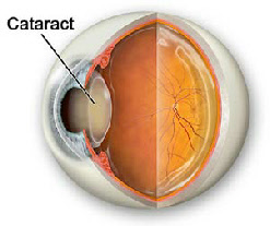
Cataracts
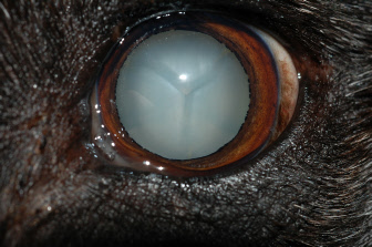
Diabetic cataract
In the normal eye, light passes through the clear cornea and lens, which act together to focus a sharp image on the retina. The retina is the nerve layer in the back of the eye that perceives the image and projects the signal to the brain for interpretation.
A cataract is an opacity of the lens. A small opacity in the lens has minimal effect on vision. When the entire lens becomes opaque, a blurred, incomplete image reaches the retina resulting in impaired vision. Dogs with advanced cataracts see light, shadows, and motion. They cannot recognize people or locate objects. Dogs with cataracts lose interest in their surroundings resulting in decreased activity.
The most common cause of cataracts in dogs is an inherited defect of the lens. In addition, dogs develop cataracts due to old age, diabetes mellitus, and other less common causes. Cataracts can also form secondary to deterioration of the retina. No treatment is beneficial for cataracts resulting from retinal degeneration since vision loss is due to damage of the nerve layer in the back of the eye. The most common cause for cataracts is cats is chronic uveitis.
The most effective treatment available to restore vision is cataract surgery. We are providing a link to the ACVO Cataract Video, which is an excellent review about the nature of cataracts and describes what is involved with cataract surgery.
Cataract surgery is routinely performed in our practice. Unless there is a problem in one of the eyes, we remove the cataract from both eyes at the same time. The success rate for restoring vision, in animals that are a good candidate for surgery, is 90 to 92%. We use a technique called phacoemulsification. With this procedure, we ultrasonically fragment and aspirate the lens through a ¼ inch incision at the periphery of the cornea. Phacoemulsification is the state-of-the-art procedure in human and veterinary medicine. As part of the surgery, we typically insert an artificial lens in the eye, to compensate for the refractive error induced by removing the lens. The procedure requires general anesthesia. We use a type of anesthesia that allows the animal to go home later the same day.